|
|

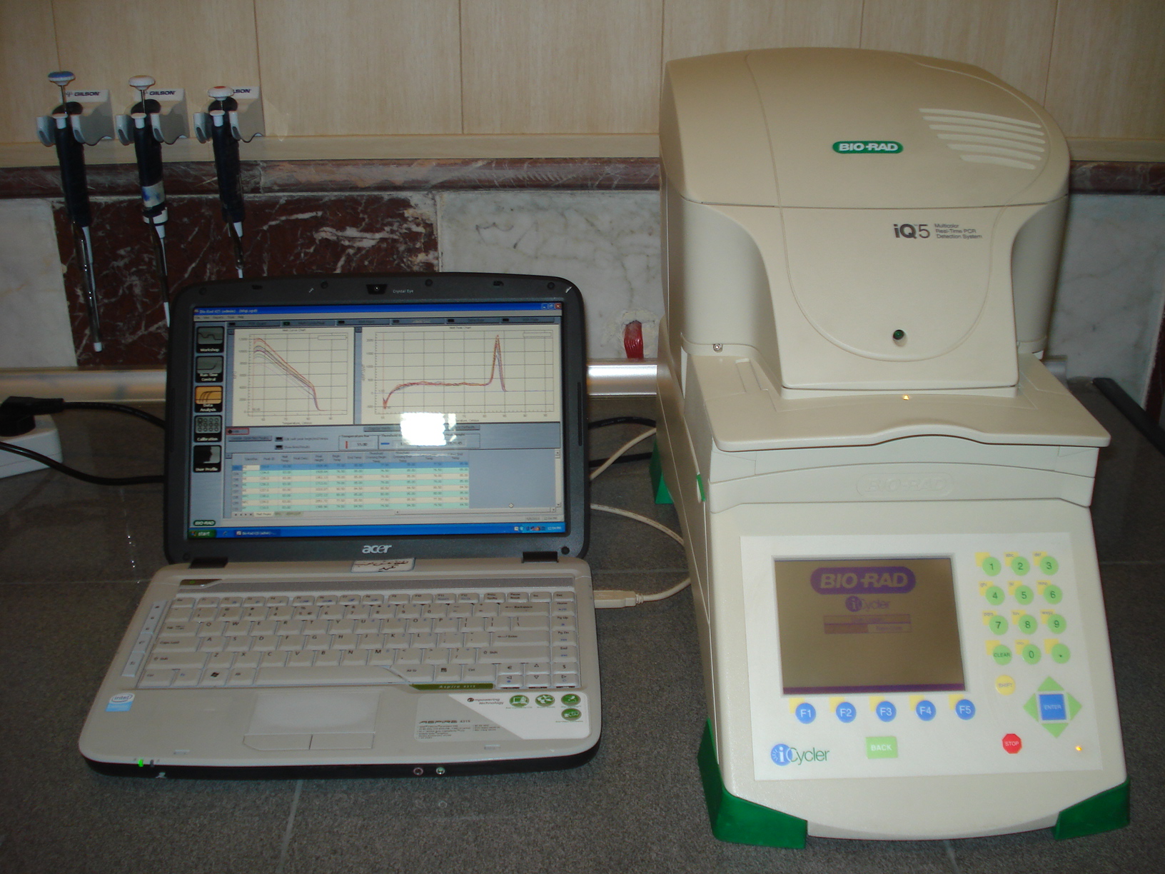
iQ5 Real Time PCR detection system
In molecular biology and biomedical research, Real Time PCR, also called quantitative real time polymerase chain reaction (qPCR) is a laboratory technique based on the PCR, which is used to amplify and simultaneously quantify a targeted nucleic acid. For one or more specific sequences, Real Time-PCR enables both detection and quantification. The quantity can be either an absolute number of copies or a relative amount when normalized to template DNA input or additional normalizing genes. There are numerous applications for real-time polymerase chain reaction in the both diagnostic and basic research:
 Gene Expression Studies Gene Expression Studies
 Chromatin Immunoprecipitation (ChIP) Chromatin Immunoprecipitation (ChIP)
 Microarray Validation Microarray Validation
 Transgene Analysis/GMO Testing Transgene Analysis/GMO Testing
 Viral/Bacterial Load Studies Viral/Bacterial Load Studies
 Allelic Discrimination/SNP Allelic Discrimination/SNP

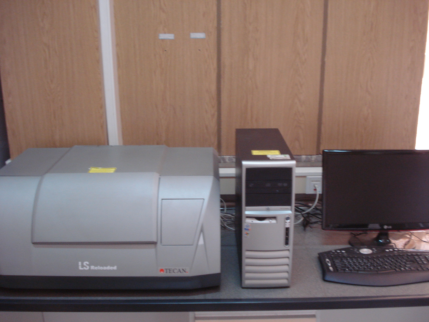
TECAN Micoarray Scanner (LS Reloaded)
A microarray is a collection of microscopic DNA spots attached to a solid surface. Scientists use DNA microarrays to measure the expression levels of large numbers of genes simultaneously or to genotype multiple regions of a genome. Each DNA spot contains 10−12 moles of a specific DNA sequence, known as probes. These can be a short section of a gene or other DNA element that are used to hybridize a cDNA or cRNA sample under high-stringency conditions. Probe-target hybridization is usually detected and quantified by detection of fluorophore, silver, or chemiluminescence-labeled targets to determine relative abundance of nucleic acid sequences in the target.

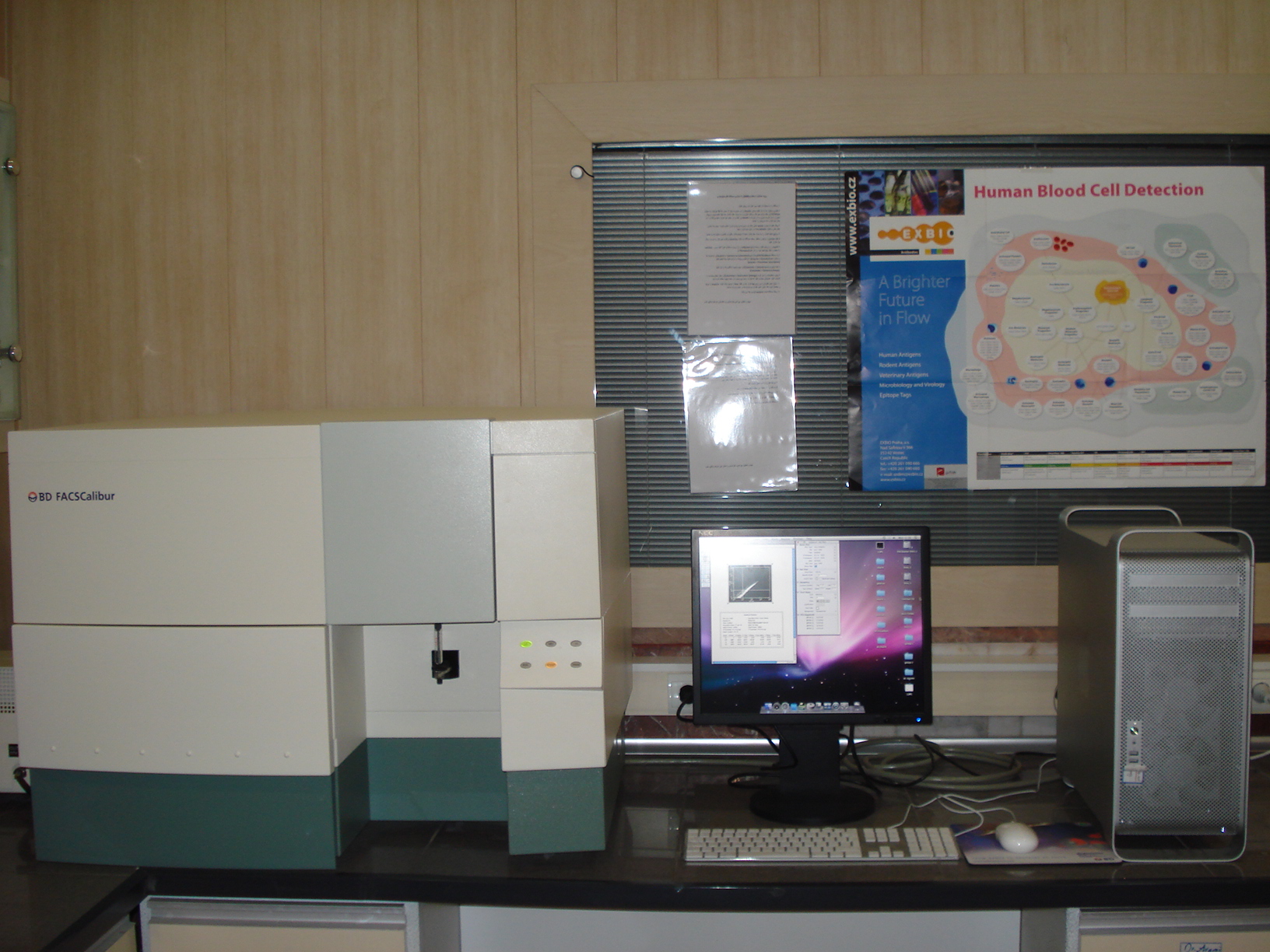
FACS flow cytometry
In cell biology, flow cytometry is a laser based, biophysical technology employed in cell counting, sorting, biomarker detection and protein engineering, by suspending cells in a stream of fluid and passing them by an electronic detection apparatus. It allows simultaneous multi-parametric analysis of the physical and/or chemical characteristics of up to thousands of particles per second.
Flow cytometry is routinely used for:
 Diagnosis of disorders, especially blood cancers Diagnosis of disorders, especially blood cancers
 Applications in basic research, clinical practice and clinical trials Applications in basic research, clinical practice and clinical trials
 A common variation is to physically sort particles based on their properties, so as to purify populations of interest A common variation is to physically sort particles based on their properties, so as to purify populations of interest

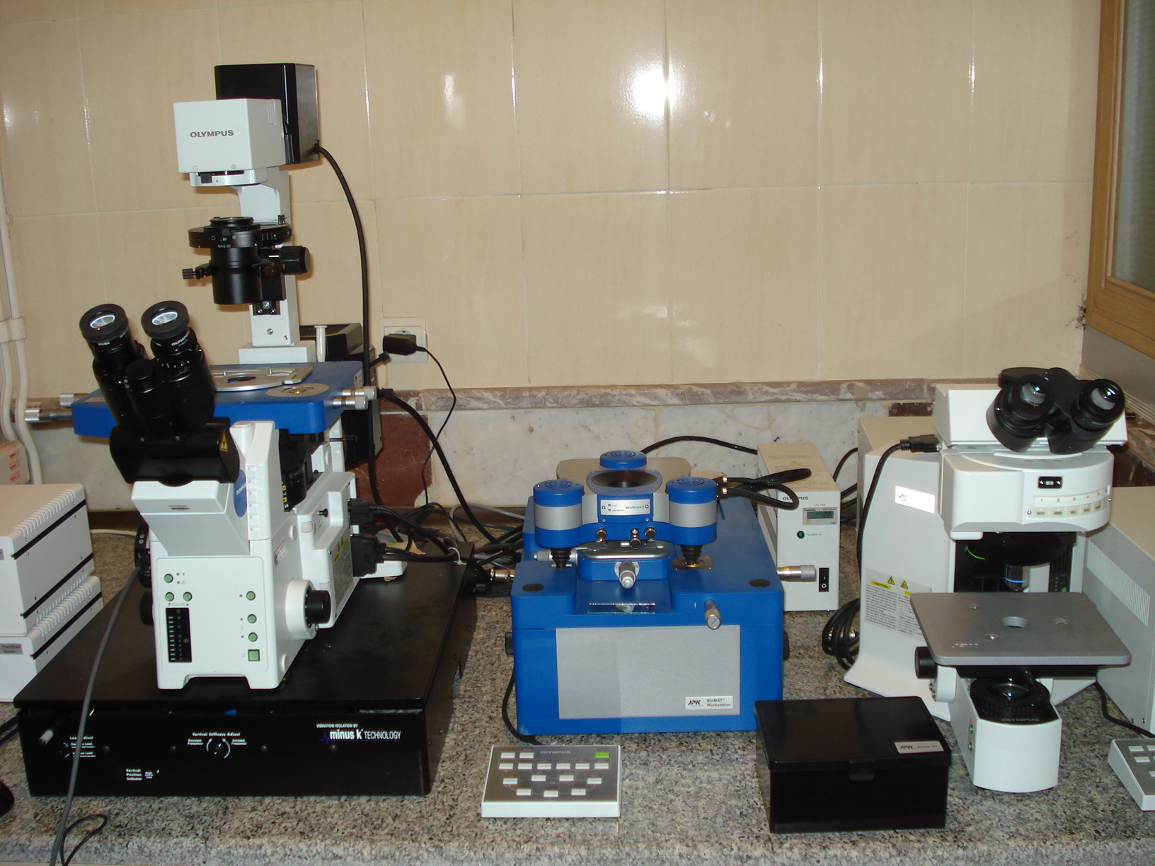
Atomic force microscopy (AFM)
The atomic force microscope (AFM) is widely used in materials science and has found many applications in biological sciences. It is a very high-resolution type of
scanning probe microscopy, with demonstrated resolution on the order of fractions of a nanometer, more than 1000 times better than the optical diffraction limit. Actually, AFM gives scientists a key tool to investigate the real world.
Applications of AFM in the biosciences include:
 analyzing the crystals of amino acids and organic monolayers analyzing the crystals of amino acids and organic monolayers
 DNA and RNA analysis, Protein-nucleic acid complexes, Chromosomes DNA and RNA analysis, Protein-nucleic acid complexes, Chromosomes
 Cellular membranes Cellular membranes
 Proteins and peptides; Molecular crystals Proteins and peptides; Molecular crystals
 Ligand-receptor binding Ligand-receptor binding
 Polymers and biomaterials Polymers and biomaterials

Scanning tunneling microscope (STM)
The STM is based on the concept of quantum tunneling. When a conducting tip is brought very near to the surface to be examined, a bias (voltage difference) applied between the two can allow electrons to tunnel through the vacuum between them. A scanning tunneling microscope (STM) is an instrument for imaging surfaces at the atomic level. For an STM, good resolution is considered to be 0.1 nm lateral resolution and 0.01 nm depth resolution.
Applications of AFM in the biosciences include:
 The STM works best with semiconductors, metals on semiconductors, metal on metals including surface alloys, oxygen on metals, molecules adsorbed on metals The STM works best with semiconductors, metals on semiconductors, metal on metals including surface alloys, oxygen on metals, molecules adsorbed on metals
 Study of fixed organic molecules surface, such as DNA molecules Study of fixed organic molecules surface, such as DNA molecules
Small Animal Imaging System
Small animal Imaging Systems provides high performance optical molecular imaging of near-IR fluorescence, isotopic, and luminescence labels in small animals. In vivo Imaging system:
 Enhances anatomical localization of molecular imaging agent signals in small animals, organs & tissues Enhances anatomical localization of molecular imaging agent signals in small animals, organs & tissues
 Enables accurate quantitation of biomolecules of interest in basic research, drug discovery, drug development, and therapeutic monitoring applications utilizing small animals Enables accurate quantitation of biomolecules of interest in basic research, drug discovery, drug development, and therapeutic monitoring applications utilizing small animals
 Allows simple switching between multiple imaging modalities including near-IR fluorescence, multiwavelength fluorescence, luminescence, radioisotopic – without disturbing the sample Allows simple switching between multiple imaging modalities including near-IR fluorescence, multiwavelength fluorescence, luminescence, radioisotopic – without disturbing the sample
Versatile – sample chamber also accommodates in vitro assay formats including blots, plates & gels
|
|
|
|
|
|
|